
Page 404 - The Vasculitides, Volume 1: General Considerations and Systemic Vasculitis
P. 404
378 Rami N. Al-Rohil and J. Andew Carlson
Circumstantial evidence of vessel wall damage includes lamination of the adventitia, media
and/or intima of vessels or so-called onion skinning (Figure 7); perivascular nuclear dust or
leukocytoclasia (Figure 8) without fibrin deposits such as in early evolving LCV; sharply
defined loss of the elastic lamina associated with acellular scar tissue in the healed stage of
muscular vessel vasculitis; and subendothelial, intramuscular, and adventitial inflammatory
cells. Neovascularization of the adventitia (Figure 9) and the formation of small capillaries
are prominent features of mature and older lesions in chronic localized SVV such as erythema
elevatum dinutum, medium vessel vasculitides (MVV) such as polyarteritis nodosa (PAN),
and large vessel vasculitides (LVV) such as giant cell arteritis (GCA), Luminal obliteration or
endarteritis obliterans, the ischemic consequence of lymphocytic and granulomatous
vasculitides, affects small- to medium-sized arteries.
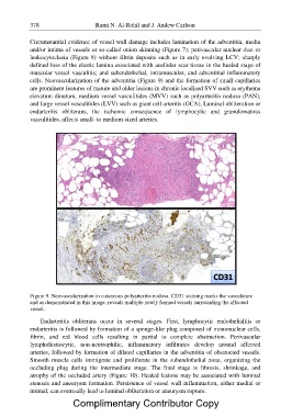
Figure 9. Neovascularization in cutaneous polyarteritis nodosa. CD31 staining marks the vasculature
and as demonstrated in this image, reveals multiple newly formed vessels surrounding the affected
vessel.
Endarteritis obliterans occur in several stages. First, lymphocytic endothelialitis or
endarteritis is followed by formation of a sponge-like plug composed of mononuclear cells,
fibrin, and red blood cells resulting in partial to complete obstruction. Perivascular
lymphohistiocytic, non-neutrophilic, inflammatory infiltrates develop around affected
arteries, followed by formation of dilated capillaries in the adventitia of obstructed vessels.
Smooth muscle cells immigrate and proliferate in the subendothelial zone, organizing the
occluding plug during the intermediate stage. The final stage is fibrosis, shrinkage, and
atrophy of the occluded artery (Figure 10). Healed lesions may be associated with luminal
stenosis and aneurysm formation. Persistence of vessel wall inflammation, either medial or
intimal, can eventually lead to luminal obliteration or aneurysm rupture.
Complimentary Contributor Copy