
Page 403 - The Vasculitides, Volume 1: General Considerations and Systemic Vasculitis
P. 403
Dermatologic Aspects of Systemic Vasculitis 377
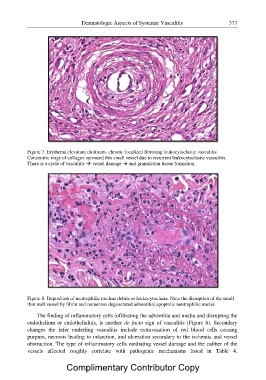
Figure 7. Erythema elevatum diutinum- chronic localized fibrosing leukocytoclastic vasculitis.
Concentric rings of collagen surround this small vessel due to recurrent leukocytoclastic vasculitis.
There is a cycle of vasculitis ? vessel damage ? and granulation tissue formation.
Figure 8. Deposition of neutrophilic nuclear debris or leukocytoclasia. Note the disruption of the small
thin wall vessel by fibrin and numerous degenerated adventitial apoptotic neutrophilic nuclei.
The finding of inflammatory cells infiltrating the adventitia and media and disrupting the
endothelium or endothelialitis, is another de facto sign of vasculitis (Figure 6). Secondary
changes the infer underling vasculitis include extravasation of red blood cells causing
purpura, necrosis leading to infarction, and ulceration secondary to the ischemia and vessel
obstruction. The type of inflammatory cells mediating vessel damage and the caliber of the
vessels affected roughly correlate with pathogenic mechanisms listed in Table 4.
Complimentary Contributor Copy