Page 402 - The Vasculitides, Volume 1: General Considerations and Systemic Vasculitis
P. 402
376 Rami N. Al-Rohil and J. Andew Carlson

             Table 3. Histologic Diagnostic Criteria for Cutaneous Vasculitis and
                                          Associated Histology1

  Histologic signs of acute (active) vasculitis
  Dermal small vessels (venules and arterioles) (2 of 3* criteria needed)
  *Angiocentric(†) and/or angio-invasive inflammatory infiltrates
  *Disruption and/or destruction of vessel wall by inflammatory infiltrate
  *Intramural and/or intraluminal fibrin deposition (“fibrinoid necrosis”)
  Dermal-Subcutaneous muscular vessels (small arteries and veins)
  *Infiltration of muscular vessel wall by inflammatory cells
  *Intramural and/or intraluminal fibrin deposition (“fibrinoid necrosis”)§
  †Secondary changes of active vasculitis (suggestive of, but not diagnostic of vasculitis)
  RBC extravasation (petechiae, purpura, hematoma)
  Nuclear dust, perivascular (leukocytoclasia)
  Endothelial swelling, sloughing or necrosis
  Eccrine gland necrosis (or regeneration with basal cell hyperplasia)
  Ulceration
  Necrosis/infarction
  Histologic sequela of vasculitis (chronic signs and healed lesions of vasculitis)
  †Lamination (onion-skinning) of vessel wall constituents
  †Luminal obliteration (endarteritis obliterans)
  *Segmental or complete loss of elastic lamina in medium and large vessels associated with acellular scar
  tissue
  †Reactive angioendotheliomatosis
  Neo-vascularization of adventitia
*: Required for diagnosis of vasculitis. §: Intraluminal fibrin deposition can be found in nonvasculitic

     arterial lesions such as malignant hypertension and anti-phospholipid antibody syndrome. †: Other
     types of vessel injury (pseudovasculitis (91)) can cause same pattern. 1 Adapted from [1].

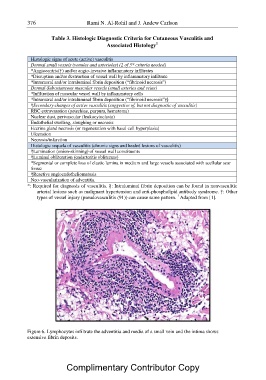
Figure 6. Lymphocytes infiltrate the adventitia and media of a small vein and the intima shows
extensive fibrin deposits.

            Complimentary Contributor Copy

