
Page 397 - The Vasculitides, Volume 1: General Considerations and Systemic Vasculitis
P. 397
Dermatologic Aspects of Systemic Vasculitis 371
by forming capillary loops. These loops are composed of terminal arteriole, capillaries
(arterial and venous), and post-capillary venules (Figure 1).
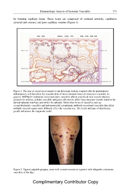
Figure 1. The size of vessel involvement is one histologic feature coupled with the predominant
inflammatory cell that allow for classification of most common forms of cutaneous vasculitis. In
general, HSP/IgAV cutaneous leukocytoclastic vasculitis affects superficial skin vessels whereas
polyarteritis nodosa, nodular vasculitis and giant cell arteritis affect deep muscular vessels found at the
dermal-subcutis interface and within the subcutis. Most other forms of vasculitis such as
cryoglobulinemic vasculitis and anti-neutrophil cytoplasmic antibody-associated vasculitis that affect
multiple visceral organs more diffusely affect the vascular tree. The depth and type of skin biopsy
greatly influences the diagnostic yield.
Figure 2. Typical palpable purpura, some with central necrosis in a patient with idiopathic cutaneous
vasculitis of the legs.
Complimentary Contributor Copy