Page 396 - The Vasculitides, Volume 1: General Considerations and Systemic Vasculitis
P. 396
370 Rami N. Al-Rohil and J. Andew Carlson

any trigger or associated systemic disease, known as idiopathic cutaneous leukocytoclastic
vasculitis (LCV) [1]. Cutaneous vasculitis manifests as urticaria, erythema, petechiae,
purpura, purpuric papules, hemorrhagic vesicles and bullae, nodules, livedo racemosa, deep
punched out ulcers and digital gangrene [2]. Skin biopsy and dermatopathology contribute
relevant information however they require correlation with the clinical history, physical exam
and laboratory findings in order to reach an accurate diagnosis in a given affected patient. The
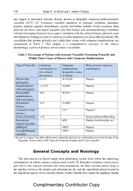
vasculitides that present primarily and within their course with cutaneous manifestations are
summarized in Table 1. This chapter is a comprehensive overview of the clinical
dermatologic aspects of primary and secondary vasculitides.

    Table 1. Percentage of Patients with Systemic Vasculitis Presenting Primarily and
                Within Their Course of Disease with Cutaneous Manifestations

Type of Vasculitis Cutaneous        Cutaneous            Most common cutaneous

                    manifestation as manifestations      manifestation

                    first clinical  during their course

                    presentation    of disease

Microscopic         4-14%           44-62.4%             Purpura

polyangiitis

Granulomatosis <1-21%               10-40%               Purpura

with polyangiitis

Eosinophilic        14%             40-81%               Purpura

granulomatosis

with polyangiitis

Polyarteritis       11%             25-60%               Purpura

Nodosa

Connective tissue -                 12% Purpura*

diseases

Giant cell arteritis <1%            -                    Scalp tenderness/blanching

Levamisole-         -               60% Purpura involving the ears

induced vasculitis

HSP/IgAV            -               100%                 Purpura

Cryoglobulinemic -                  100%                 Purpura

vasculitis

* Except for Lupus Vasculitis which most commonly presents as erythematous punctate lesions of the
     fingertips. Abbreviation: HSP, Henoch-Schönlein Purpura; IgAV, IgA vasculitis.

               General Concepts and Nosology

     The skin receives its blood supply from penetrating vessels from within the underlying
subcutaneous fat which contains medium sized vessels [3]. Branches of medium-sized vessels
give rise to two vascular plexuses that intercommunicate, the deep vascular plexus lying at
the interface between the dermis and subcutaneous fat, and the superficial plexus located in
the superficial aspects of the reticular dermis. Further distally they supply the papillary dermis

                Complimentary Contributor Copy

