
Page 411 - The Vasculitides, Volume 1: General Considerations and Systemic Vasculitis
P. 411
Dermatologic Aspects of Systemic Vasculitis 385
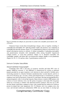
Figure 16. Multiple red collagen lytic granulomas in a patient with eosinophilic granulomatosis with
polyangiitis.
Cutaneous biopsy reveals three histopathologic changes, alone or together, including 1)
eosinophil-rich neutrophilic SVV affecting dermal venules and arterioles, less commonly an
eosinophil-rich muscular arteritis (Figure 15) or granulomatous histiocyte-rich arteritis of the
dermal subcutaneous junction or subcutis; 2) dermal eosinophilia; 2) palisading neutrophilic
and granulomatous dermatitis with neutrophilic debris among basophilic degenerated
collagen bundles, so called blue granuloma (Figure 13) or abundant eosinophils and
eosinophilic granules and debris that coat degenerated collagen bundles or red granulomas
(Figure 16) [18, 33, 34]; and less often, 3) granulomatous arteritis [34].
Immune Complex Vasculitides
Henoch Schönlein Purpura/IgAV
Comprising about 10% of all cases of cutaneous vasculitis and about 90% cases of
primary childhood vasculitis [1, 18], HSP/IgAV is characterized by retiform or patterned
purpura preceded by an upper respiratory tract infection in about one-half of children and a
recent drug exposure in a minority. Isolated or predominate IgA vascular deposits, and two or
more of the following: age ? 20 years, colicky gastrointestinal pain or hematochezia, an upper
respiratory tract infection prodrome, hematuria or renal biopsy showing mesangioproliferative
glomerulonephritis with or without IgA deposits. Renal disease is found in HSP patients with
a history of recent infection, fever, and the spread of purpura to the trunk [35]. Long-term
follow-up is necessary in children with abnormal baseline urinalysis [36] since up to 20%
develop chronic renal failure compared to those with a normal baseline urinalysis in which no
person developed later renal impairment [36, 37]. Patients with fever, purpura above the
waist, and elevated erythrocyte sedimentation (ESR) rate are more likely to develop IgA
Complimentary Contributor Copy