
Page 414 - The Vasculitides, Volume 1: General Considerations and Systemic Vasculitis
P. 414
388 Rami N. Al-Rohil and J. Andew Carlson
ischemic endarteritis obliterans, and systemic features of malaise, weight loss, and fever due
to release of inflammatory cytokines. The cutaneous signs consist of scalp tenderness;
blanching, decreased or loss of temporal artery pulses, and cord-like temporal artery
thickening. Accurate and timely diagnosis is important because serious morbidity especially
loss of vision if the correct treatment with corticosteroids are delayed. Skin biopsies that
include muscular vessels of the subcutis and temporal artery biopsies show granulomatous
vasculitis with giant-cell-containing inflammatory infiltrates, while the essential diagnostic
features of temporal artery biopsy include segmental inflammation with disruption of media
and intima, and fragmentation of the internal elastic lamina.
Secondary Vasulitides
Connective Tissue Diseases
Lupus Vasculitis
Ramos-Casals and colleagues [9] noted cutaneous vasculitis in 76% of 670 patients with
SLE. The commonest cutaneous lesions were erythematous punctate lesions of the fingertips
and palms, followed by purpura. Female SLE patients, anti-Ro seropositive, had a 1.63
greater risk of develop cutaneous vasculitis [56].
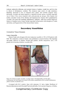
Figure 18. Cutaneous Lupus vasculitis. A young woman with longstanding systemic lupus
erythematosus and multiorgan involvement with nodules, pyodermatous ulcers, painful erythematous
palmar macules and punctate scars.
Compared with SLE controls, those with cutaneous LV had a higher likelihood of
Raynaud phenomenon and ribosomal P protein antibodies, but no greater frequency of kidney
Complimentary Contributor Copy