
Page 415 - The Vasculitides, Volume 1: General Considerations and Systemic Vasculitis
P. 415
Dermatologic Aspects of Systemic Vasculitis 389
or nervous system involvement [57]. Patients with LV patients typically present with livedo
reticularis, anemia, high ESR, and anti-La/SS-B antibodies than SLE patients without
vasculitis. Arterioles and post-capillary venules are the most commonly affected by vasculitis,
manifested as purpura, vesiculobullous lesions, urticaria, and splinter hemorrhages. Arterial
vessel involvement and multiorgan involvement is suggested by cutaneous ulcers, nodules,
digital gangrene, necrotizing livedo reticularis, punctate acral scars, and pyoderma
gangrenosum (PG)-like lesions (Figure 18). There may be p-ANCA and less often c-ANCA-
seropositivity. Skin biopsy shows neutrophilic vasculitis with lesions that can resemble either
typical LCV or PAN, or coexistence of both small and muscular vessel vasculitis in the same
biopsy specimen.
Rheumatoid Vasculitis
The extra-articular ExRA) manifestation of clinically-evident RAV were noted in 2% of
patients [17] compared to 15% to 31% in postmortem studied cases of RA [58]. RAV
typically affects small- and medium size blood vessels and is associated with increased
premature mortality with mortality in 40 % of patients by 5 years as well as significant
morbidity due to both organ damage from vasculitis and consequences of the cytotoxic
treatment [59]. Cutaneous manifestations of RAV are graded mild, moderate, and severe.
Mild cutaneous RAV presents with nail-fold telangiectasia, thrombosis, minute digital
ulceration, petechiae, and livedo reticularis. Moderate cases manifest palpable purpura.
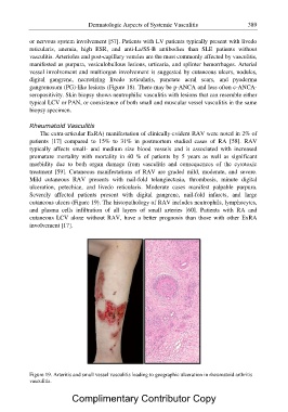
Severely affected patients present with digital gangrene, nail-fold infarcts, and large
cutaneous ulcers (Figure 19). The histopathology of RAV includes neutrophils, lymphocytes,
and plasma cells infiltration of all layers of small arteries [60]. Patients with RA and
cutaneous LCV alone without RAV, have a better prognosis than those with other ExRA
involvement [17].
Figure 19. Arteritis and small vessel vasculitis leading to geographic ulceration in rheumatoid arthritis
vasculitis.
Complimentary Contributor Copy