
Page 305 - The Vasculitides, Volume 1: General Considerations and Systemic Vasculitis
P. 305
Systemic Vasculitis of the Gastrointestinal Tract 279
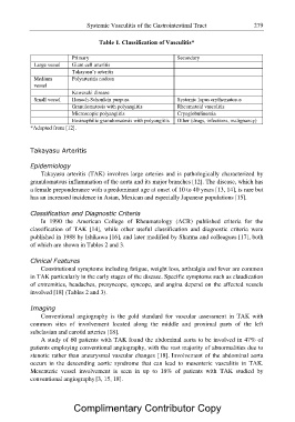
Table 1. Classification of Vasculitis*
Primary Secondary
Large vessel Giant cell arteritis Systemic lupus erythematosus
Takayasu’s arteritis Rheumatoid vasculitis
Cryoglobulinemia
Medium Polyarteritis nodosa Other (drugs, infections, malignancy)
vessel
Kawasaki disease
Small vessel Henoch-Schonlein purpura
Granulomatosis with polyangiitis
Microscopic polyangiitis
Eosinophilic granulomatosis with polyangiitis
*Adapted from [12].
Takayasu Arteritis
Epidemiology
Takayasu arteritis (TAK) involves large arteries and is pathologically characterized by
granulomatous inflammation of the aorta and its major branches [12]. The disease, which has
a female preponderance with a predominant age at onset of 10 to 40 years [13, 14], is rare but
has an increased incidence in Asian, Mexican and especially Japanese populations [15].
Classification and Diagnostic Criteria
In 1990 the American College of Rheumatology (ACR) published criteria for the
classification of TAK [14], while other useful classification and diagnostic criteria were
published in 1988 by Ishikawa [16], and later modified by Sharma and colleagues [17], both
of which are shown in Tables 2 and 3.
Clinical Features
Constitutional symptoms including fatigue, weight loss, arthralgia and fever are common
in TAK particularly in the early stages of the disease. Specific symptoms such as claudication
of extremities, headaches, presyncope, syncope, and angina depend on the affected vessels
involved [18] (Tables 2 and 3).
Imaging
Conventional angiography is the gold standard for vascular assessment in TAK with
common sites of involvement located along the middle and proximal parts of the left
subclavian and carotid arteries [18].
A study of 60 patients with TAK found the abdominal aorta to be involved in 47% of
patients employing conventional angiography, with the vast majority of abnormalities due to
stenotic rather than aneurysmal vascular changes [18]. Involvement of the abdominal aorta
occurs in the descending aortic syndrome that can lead to mesenteric vasculitis in TAK.
Mesenteric vessel involvement is seen in up to 18% of patients with TAK studied by
conventional angiography [3, 15, 18].
Complimentary Contributor Copy