
Page 306 - The Vasculitides, Volume 1: General Considerations and Systemic Vasculitis
P. 306
280 Dimitri Chanouzas and Matthew David Morgan
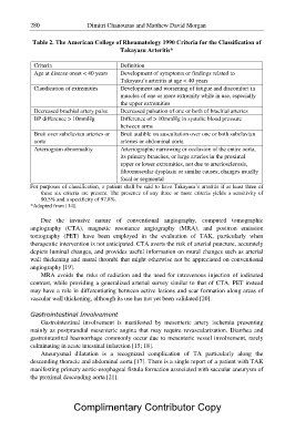
Table 2. The American College of Rheumatology 1990 Criteria for the Classification of
Takayasu Arteritis*
Criteria Definition
Age at disease onset < 40 years Development of symptoms or findings related to
Takayasu’s arteritis at age < 40 years
Claudication of extremities Development and worsening of fatigue and discomfort in
muscles of one or more extremity while in use, especially
the upper extremities
Decreased brachial artery pulse Decreased pulsation of one or both of brachial arteries
BP difference > 10mmHg Difference of > 10mmHg in systolic blood pressure
between arms
Bruit over subclavian arteries or Bruit audible on auscultation over one or both subclavian
aorta arteries or abdominal aorta
Arteriogram abnormality Arteriographic narrowing or occlusion of the entire aorta,
its primary branches, or large arteries in the proximal
upper or lower extremities, not due to arteriosclerosis,
fibromuscular dysplasia or similar causes; changes usually
focal or segmental
For purposes of classification, a patient shall be said to have Takayasu’s arteritis if at least three of
these six criteria are present. The presence of any three or more criteria yields a sensitivity of
90.5% and a specificity of 97.8%.
*Adapted from [14].
Due the invasive nature of conventional angiography, computed tomographic
angiography (CTA), magnetic resonance angiography (MRA), and positron emission
tomography (PET) have been employed in the evaluation of TAK, particularly when
therapeutic intervention is not anticipated. CTA averts the risk of arterial puncture, accurately
depicts luminal changes, and provides useful information on mural changes such as arterial
wall thickening and mural thrombi that might otherwise not be appreciated on conventional
angiography [19].
MRA avoids the risks of radiation and the need for intravenous injection of iodinated
contrast, while providing a generalized arterial survey similar to that of CTA. PET instead
may have a role in differentiating between active lesions and scar formation along areas of
vascular wall thickening, although its use has not yet been validated [20].
Gastrointestinal Involvement
Gastrointestinal involvement is manifested by mesenteric artery ischemia presenting
mainly as postprandial mesenteric angina that may require revascularization. Diarrhea and
gastrointestinal haemorrhage commonly occur due to mesenteric vessel involvement, rarely
culminating in acute intestinal infarction [15; 18].
Aneurysmal dilatation is a recognized complication of TA particularly along the
descending thoracic and abdominal aorta [17]. There is a single report of a patient with TAK
manifesting primary aortic-esophageal fistula formation associated with saccular aneurysm of
the proximal descending aorta [21].
Complimentary Contributor Copy