
Page 307 - The Vasculitides, Volume 1: General Considerations and Systemic Vasculitis
P. 307
Systemic Vasculitis of the Gastrointestinal Tract 281
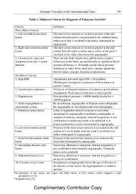
Table 3. Ishikawa Criteria for Diagnosis of Takayasu Arteritis*
Criteria Definition
Three Major Criteria
1. Left mid subclavian artery The most severe stenosis or occlusion present in the mid
lesion portion from the point 1 cm proximal to the vertebral artery
orifice up to that 3 cm distal to the orifice determined by
angiography
2. Right mid subclavian artery The most severe stenosis or occlusion present in the mid
lesion portion from the right vertebral artery orifice to the point 3
cm distal to the orifice determined by angiography
3. Characteristic signs and These include limb claudication, pulselessness or pulse
symptoms of at least 1 month differences in the limbs, an unobtainable or significant blood
duration pressure difference (> 10 mmHg systolic blood pressure
difference in limb), fever, neck pain, transient amaurosis,
blurred vision, syncope, dyspnea or palpitations
Ten Minor Criteria
1. High ESR Unexplained persistent high ESR > 20 mm/hour
(Westergren) at diagnosis or presence of the evidence in
patient’s history
2. Carotid artery tenderness Unilateral or bilateral tenderness of common carotid arteries
on palpation. Neck muscle tenderness is unacceptable
3. Hypertension Persistent blood pressure > 140/90 mmHg brachial or >
160/90 popliteal
4. Aortic regurgitation or By auscultation, angiography or Doppler echocardiography.
annuloaortic ectasia By angiography or two-dimensional echocardiography
5. Pulmonary artery lesion Lobar or segmental arterial occlusion or equivalent
determined by angiography or perfusion scintigraphy, or
presence of stenosis, aneurysm, luminal irregularity or any
combination in pulmonary trunk or in unilateral or in
bilateral pulmonary arteries determined by angiography
6. Left mid common carotid Presence of the most severe stenosis or occlusion in the mid
lesion portion of 5 cm in length from the point 2 cm distal to its
orifice determined by angiography
7. Distal brachiocephalic trunk Presence of the most severe stenosis or occlusion in the
lesion distal third determined by angiography
8. Descending thoracic aorta Narrowing, dilatation or aneurysm, luminal irregularity or
lesion any combination determined by angiography. Tortuosity
alone is not acceptable
9. Abdominal aorta lesion Narrowing, dilatation or aneurysm, luminal irregularity or
aneurysm combination
10. Coronary artery lesion Documented on angiography below the age of 30 years in
the absence of risk factors like hyperlipidemia or diabetes
mellitus
The presence of two major, one major and three minor, or four minor criteria suggests a high
probability of TA.
*Adapted from [17].
Complimentary Contributor Copy