
Page 162 - The Vasculitides, Volume 1: General Considerations and Systemic Vasculitis
P. 162
138 J. Charles Jennette, Ronald J. Falk and Adil. H. M. Gasim
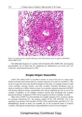
Figure 10. Leukocytoclastic angiitis affecting a small subcutaneous artery in a patient with Beches
disease (H&E stain).
The differential diagnosis of a patient with CS includes GPA, EGPA, RV, and relapsing
polychondritis, all of which may be complicated by inflammatory eye disease and, less
commonly, sensorineural hearing loss [30].
Single-Organ Vasculitis
CHCC 2012 defines SOV as vasculitis in arteries or veins of any size in a single organ
that has no features that indicate that it is a limited expression of a systemic vasculitis [1]. The
involved organ and vessel type should be included in the name, for example cutaneous small
vessel vasculitis, testicular arteritis, and CNS vasculitis. Vasculitis distribution may be
unifocal, multifocal or diffuse within an organ. Some patients originally diagnosed with SOV
will develop additional disease manifestations that warrant redefining the case as one of the
systemic vasculitides, as for example cutaneous arteritis later developing into systemic
polyarteritis nodosa. A diagnosis of SOV always requires exclusion of systemic vasculitis.
Many forms of SOV have been reported, for example cutaneous LCV, cutaneous arteritis
or cutaneous polyarteritis [33, 34], primary angiitis of the CNS (PACNS) [35] or the
equivalent term primary CNS vasculitis (PCNSV) [36]; isolated aortitis, and others [37]. The
histopathologic pattern of injury can resemble any of the recognized forms of systemic
vasculitis, although necrotizing arteries and granulomatous arteritis are most frequent.
Complimentary Contributor Copy