Page 160 - The Vasculitides, Volume 1: General Considerations and Systemic Vasculitis
P. 160
136 J. Charles Jennette, Ronald J. Falk and Adil. H. M. Gasim

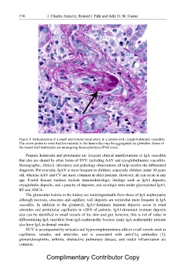
Figure 9. Inflammation of a small interlobular renal artery in a patient with cryoglobulinemic vasculitis.
The arrow points to some hyaline material in the lumen that may be aggregated cry globulins. Some of
the vessel wall leukocytes are undergoing leukocytoclasia (PAS stain).

     Purpura hematuria and proteinuria are frequent clinical manifestations of IgA vasculitis
that also are shared by other forms of SVV, including AAV and cryoglobulinemic vasculitis.
Demographic, clinical, laboratory and pathology observations all help resolve the differential
diagnosis. For example, IgAV is most frequent in children, especially children under 10 years
old, whereas AAV and CV are more common in older patients. However, all can occur at any
age. Useful disease markers include immunohistologic findings such as IgA1 deposits,
cryoglobulin deposits, and a paucity of deposits; and serologic tests under glycosylated IgA1,
RF and ANCA.

     The glomerular lesions in the kidney are indistinguishable from those of IgA nephropathy
although necrosis, crescents and capillary wall deposits are somewhat more frequent in IgA
vasculitis. In addition to the glomeruli, IgA1-dominant immune deposits occur in renal
arterioles and peritubular capillaries in <20% of patients. IgA1-dominant immune deposits
also can be identified in small vessels of the skin and gut; however, this is not of value in
differentiating IgA vasculitis from IgA nephropathy because many IgA nephropathy patients
also have IgA in dermal venules.

     HUV is accompanied by urticaria and hypocomplementemia affects small vessels such as
capillaries, venules, and arterioles, and is associated with anti-C1q antibodies [1];
glomerulonephritis, arthritis, obstructive pulmonary disease, and ocular inflammation are
common.

            Complimentary Contributor Copy

