
Page 157 - The Vasculitides, Volume 1: General Considerations and Systemic Vasculitis
P. 157
Nomenclature and Pathologic Features of Vasculitides 133
ab
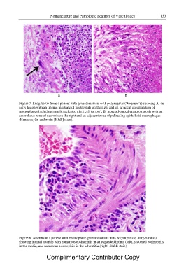
Figure 7. Lung tissue from a patient with granulomatosis with polyangiitis (Wegener?s) showing A: an
early lesion with an intense infiltrate of neutrophils on the right and an adjacent accumulation of
macrophages including a multinucleated giant cell (arrow), B: more advanced granulomatosis with an
amorphous zone of necrosis on the right and an adjacent zone of palisading epithelioid macrophages
(Hematoxylin and eosin [H&E] stain).
Figure 8. Arteritis in a patient with eosinophilic granulomatosis with polyangiitis (Churg-Strauss)
showing intimal arteritis with numerous eosinophils in an expanded intima (left), scattered eosinophils
in the media, and numerous eosinophils in the adventitia (right) (H&E stain).
Complimentary Contributor Copy