
Page 152 - The Vasculitides, Volume 1: General Considerations and Systemic Vasculitis
P. 152
128 J. Charles Jennette, Ronald J. Falk and Adil. H. M. Gasim
ab
cd
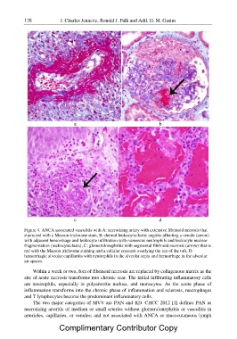
Figure 4. ANCA associated vasculitis with A: necrotizing artery with extensive fibrinoid necrosis that
stains red with a Masson trichrome stain, B: dermal leukocytoclastic angiitis affecting a venule (arrow)
with adjacent hemorrhage and leukocyte infiltration with numerous neutrophils and leukocyte nuclear
fragmentation (leukocytoclasia), C: glomerulonephritis with segmental fibrinoid necrosis (arrow) that is
red with the Masson trichrome staining and a cellular crescent overlying the top of the tuft, D:
hemorrhagic alveolar capillaritis with neutrophils in the alveolar septa and hemorrhage in the alveolar
air spaces.
Within a week or two, foci of fibrinoid necrosis are replaced by collagenous matrix as the
site of acute necrosis transforms into chronic scar. The initial infiltrating inflammatory cells
are neutrophils, especially in polyarteritis nodosa, and monocytes. As the acute phase of
inflammation transforms into the chronic phase of inflammation and sclerosis, macrophages
and T lymphocytes become the predominant inflammatory cells.
The two major categories of MVV are PAN and KD. CHCC 2012 [1] defines PAN as
necrotizing arteritis of medium or small arteries without glomerulonephritis or vasculitis in
arterioles, capillaries, or venules; and not associated with ANCA or mucocutaneous lymph
Complimentary Contributor Copy