
Page 151 - The Vasculitides, Volume 1: General Considerations and Systemic Vasculitis
P. 151
Nomenclature and Pathologic Features of Vasculitides 127
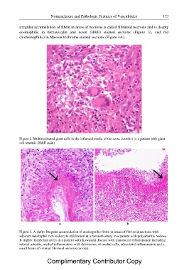
irregular accumulation of fibrin in areas of necrosis is called fibrinoid necrosis and is deeply
eosinophilic in hematoxylin and eosin (H&E) stained sections (Figure 3), and red
(fuchsinophilic) in Masson trichrome stained sections (Figure 4A).
Figure 2. Multinucleated giant cells in the inflamed media of the aorta (aortitis) in a patient with giant
cell arteritis (H&E stain).
ab
Figure 3. A (left): Irregular accumulation of eosinophilic fibrin in areas of fibrinoid necrosis with
adjacent neutrophil-rich leukocyte infiltration in a medium artery in a patient with polyarteritis nodosa.
B (right): Interlobar artery in a patient with Kawasaki disease with transmural inflammation including
intimal arteritis, medial inflammation with dehiscence of medial cells, adventitial inflammation and a
small focus of intimal fibrinoid necrosis (arrow).
Complimentary Contributor Copy