
Page 148 - The Vasculitides, Volume 1: General Considerations and Systemic Vasculitis
P. 148
124 J. Charles Jennette, Ronald J. Falk and Adil. H. M. Gasim
vessels affected since in a given patients with LVV, there may be, and often are numerically
more involved medium and small vessels than large ones.
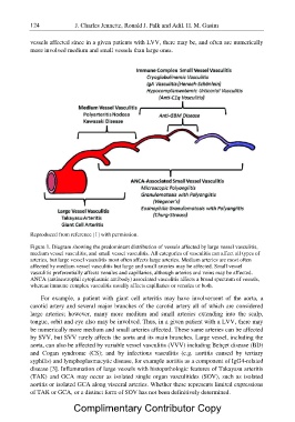
Reproduced from reference [1] with permission.
Figure 1. Diagram showing the predominant distribution of vessels affected by large vessel vasculitis,
medium vessel vasculitis, and small vessel vasculitis. All categories of vasculitis can affect all types of
arteries, but large vessel vasculitis most often affects large arteries. Medium arteries are most often
affected by medium vessel vasculitis but large and small arteries may be affected. Small vessel
vasculitis preferentially affects venules and capillaries, although arteries and veins may be affected.
ANCA (antineutrophil cytoplasmic antibody) associated vasculitis affects a broad spectrum of vessels,
whereas immune complex vasculitis usually affects capillaries or venules or both.
For example, a patient with giant cell arteritis may have involvement of the aorta, a
carotid artery and several major branches of the carotid artery all of which are considered
large arteries; however, many more medium and small arteries extending into the scalp,
tongue, orbit and eye also may be involved. Thus, in a given patient with a LVV, there may
be numerically more medium and small arteries affected. These same arteries can be affected
by SVV, but SVV rarely affects the aorta and its main branches. Large vessel, including the
aorta, can also be affected by variable vessel vasculitis (VVV) including Behçet disease (BD)
and Cogan syndrome (CS); and by infectious vasculitis (e.g. aortitis caused by tertiary
syphilis) and lymphoplasmacytic disease, for example aortitis as a component of IgG4-related
disease [3]. Inflammation of large vessels with histopathologic features of Takayasu arteritis
(TAK) and GCA may occur as isolated single organ vasculitides (SOV), such as isolated
aortitis or isolated GCA along visceral arteries. Whether these represents limited expressions
of TAK or GCA, or a distinct form of SOV has not been definitively determined.
Complimentary Contributor Copy