
Page 274 - The Vasculitides, Volume 1: General Considerations and Systemic Vasculitis
P. 274
248 Christian Pagnoux and Gerard P. Cox
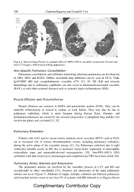
Figure 6. Massive lung fibrosis in a patient with anti-MPO-ANCA vasculitis in remission (frontal and
axial CT images), with honeycombing appearance.
Non-Specific Pulmonary Consolidation
Pulmonary consolidation and infiltrates mimicking infectious pneumonia can be observed
in GPA, MPA and EGPA. Diffuse interstitial lung infiltrates can be seen in GCA, TAK,
IgAV/HSP, BD and cryoglobulinemic vasculitis (CV) [13, 29, 30]. ILD and alveolar
hemorrhage due to pulmonary capillaritis can also occur in rheumatoid-associated vasculitis
(RAV), as with other systemic diseases such as systemic lupus erythematosus (SLE).
Pleural Effusion and Pneumothorax
Pleural effusions are common in EGPA and polyarteritis nodosa (PAN). They can be
naturally inflammatory or related to cardiac or renal failure. They may also be due to
pulmonary embolism, which is more frequent during disease flares. Pneumo- and
hydropneumothoraces are caused by the outward progression of peripheral lung nodules that
involve the pleura and cavitated [31, 32].
Pulmonary Embolism
Patients with AAV and to a lesser extent, medium-vessel vasculitis (MVV) such as PAN,
are at increased risk of venous thromboembolic events, including pulmonary embolism,
during the active phase of the vasculitic disease [33, 34]. Pulmonary embolism due to right
ventricular thrombi occurs in BD due to increased tissue-factor expression of neutrophilic
extracellular traps and neutrophil-derived microparticles [35]. Anti-PR3-ANCA GPA
antibodies with dual reactivity to plasminogen and complementary PR3 have been noted [36].
Pulmonary Artery Stenosis and Aneurysms
The pulmonary arteries are involved by the vasculitic process in LVV and BD and
exceptionally in other vasculitides [37]. Stenoses and aneurysms of the main pulmonary
arteries can occur (Figure 7). Dilations of single, multiple, unilateral and bilateral pulmonary
and bronchial arteries occur in less than 5% of patients with BD referred to as Hughes-Stovin
Complimentary Contributor Copy