
Page 270 - The Vasculitides, Volume 1: General Considerations and Systemic Vasculitis
P. 270
244 Christian Pagnoux and Gerard P. Cox
opacities on chest CT (Figure 2), and persistent bloody return of bronchioalveolar fluid during
lavage. In addition to ground-glass opacities, chest CT may reveal alveolar consolidation and
localized rather than diffuse or patchy infiltrates. CO2 diffusion capacity is usually increased
with blood in the alveoli, but pulmonary function test (PFT) is rarely indicated or performed
in patients with frank alveolar hemorrhage.
DAH is associated with renal disease in GPA, MPA and anti-GBM disease and may be
part of a pulmonary–renal syndrome. When alveolar hemorrhage occurs as an isolated event
or is associated with non-specific constitutional symptoms, diagnoses other than vasculitis
should be considered (Table 1) [14, 15]. Hemoptysis from lung capillaritis should be
differentiated from other vasculitic causes of bleeding such as submucosal vessel rupture in
patients with endobronchial GPA mucosal ulceration and rupture of larger-vessel aneurysms
in those with TAK and BD.
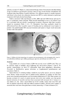
Figure 2. Diffuse alveolar hemorrhage in a patient with granulomatosis with polyangiitis (left: chest X-
ray with diffuse infiltrate; right: diffuse ground-glass opacities and infiltrates on axial CT scan).
Nodules
Lung nodules are a classical feature of GPA but can also occur in EGPA [16]. They can
be cavitated, single or multiple, and of differing size and location (Figures 3 and 4). No
specific characteristics on chest CT differentiate them from malignancy; tuberculous,
Nocardial or Burkhloderia species infections; or lymphomatoid granulomatosis and
sarcoidosis (Table 2).
Supra-infection of cavitating nodules, notably by Aspergillus species, can occur. Surgical
tissue biopsy, wedge-resection and CT-guided needle aspiration of nodules in GPA may
reveal non-caseating granulomatous and necrotizing inflammation [17-20]. Plain nodules can
become cavitated as a result of central necrosis and evolve into nodular scars that persist
beyond remission of vasculitis and have been associated with increased risk of relapse in a
few studies [21, 22]. After a mean follow-up of 26.5 months, relapse rates were 44% and
42% in patients respectively with and without persistent nodules; and non-active nodules at
remission that were more often plain than cavitated, measuring < 15 mm in diameter [21].
Complimentary Contributor Copy