
Page 113 - The Vasculitides, Volume 1: General Considerations and Systemic Vasculitis
P. 113
Experimental Autoimmune Vasculitis 89
Rat Models
A model of MPO-AAV was developed by immunization of a susceptible Wistar-Kyoto
(WKY) rat strain with human MPO, in Complete Freund?s adjuvant (CFA).
This approach resulted in generation of MPO-ANCA and MPO-reactive T-cells [17] with
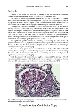
development of a crescentic, pauci-immune glomerulonephritis, and pulmonary capillaritis in
a proportion of treated animals (Figure 1) [18]. The induced anti-human MPO antibodies
cross-reacted with rat MPO, displaying significant homology to the human molecule.
Although ANCA-IgG was not found to transfer disease in this model, it did increase
leukocyte-endothelial interaction [19], and clearly showed that the composition of the
adjuvant providing the innate immune stimulation and the genetic background of the rat were
critical to the determination of severity and disease susceptibility. Lewis rats, which share the
same MHC Rt1 locus as the WKY strain, do not develop vasculitis or glomerulonephritis
despite achieving similar levels of anti-MPO antibodies, demonstrating that non-MHC genes
are important in mediating the pathogenic potential of MPO-ANCA.
The significant genetic differences conferring susceptibility to other forms of
glomerulonephritis including nephrotoxic nephritis and anti-glomerular basement membrane
disease, in these rats has been described, largely related to differences in macrophage
reactivity and Fc receptor activation [20].
The role of certain genetic loci as susceptibility factors in the MPO-AAV model and
resulting disease severity was examined using congenic Lewis/WKY animals demonstrating
the critical role of chromosome 13 and 16 susceptibility loci in the development of
experimental autoimmune vasculitis [21].
While the murine T-cell mediated model implicates deposited MPO directing a delayed
type hypersensitivity response within the kidney, it does not appear to be the main mechanism
in the rat MPO autoimmune model [18] as there is no evidence of deposited MPO.
Figure 1. Photomicrograph of a single glomerulus from a rat with experimental autoimmune vasculitis
demonstrating crescentic change (PAS x400).
Complimentary Contributor Copy