
Page 345 - The Vasculitides, Volume 1: General Considerations and Systemic Vasculitis
P. 345
Giant Cell Arteritis 319
in TAB analysis [55, 99]. The side selected for TAB should be the one, if present, with
lateralizing symptoms or signs [102].
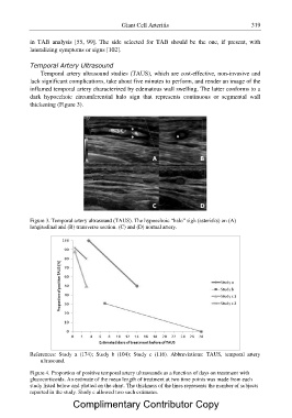
Temporal Artery Ultrasound
Temporal artery ultrasound studies (TAUS), which are cost-effective, non-invasive and
lack significant complications, take about five minutes to perform, and render an image of the
inflamed temporal artery characterized by edematous wall swelling. The latter conforms to a
dark hypoechoic circumferential halo sign that represents continuous or segmental wall
thickening (Figure 3).
Figure 3. Temporal artery ultrasound (TAUS). The hypoechoic “halo” sigh (asterisks) on (A)
longitudinal and (B) transverse section. (C) and (D) normal artery.
References: Study a (174); Study b (104); Study c (116). Abbreviations: TAUS, temporal artery
ultrasound.
Figure 4. Proportion of positive temporal artery ultrasounds as a function of days on treatment with
glucocorticoids. An estimate of the mean length of treatment at two time points was made from each
study listed below and plotted on the chart. The thickness of the lines represents the number of subjects
reported in the study. Study c allowed two such estimates.
Complimentary Contributor Copy