
Page 339 - The Vasculitides, Volume 1: General Considerations and Systemic Vasculitis
P. 339
Giant Cell Arteritis 313
composed of CD4+ T-cells and macrophages located at the intima-media junction near
fragments of the internal elastic lamina. Other TAB specimens manifest lympho-
mononuclear-predominant panarteritis with occasional neutrophils and eosinophils without
giant cells [29].
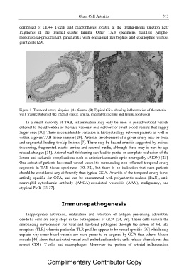
Figure 1. Temporal artery biopsies. (A) Normal (B) Typical GSA showing inflammation of the arterial
wall, fragmentation of the internal elastic lamina, internal thickening and luminal occlusion.
In a small minority of TAB, inflammation may only be seen in periadventitial vessels
external to the adventitia or the vasa vasorum in a network of small blood vessels that supply
larger ones [30]. There is considerable variation in histopathology between patients as well as
within a given TAB tissue sample [29]. Arteritic involvement of a given artery may be focal
and segmental leading to skip lesions [7]. There may be healed arteritis suggested by intimal
thickening, fragmented elastic lamina and scarred media, although these may in part be age
related changes [31]. Arterial wall thickening can lead to partial or complete occlusion of the
lumen and ischemic complications such as anterior ischaemic optic neuropathy (AION) [23].
One subset of patients has small-vessel vasculitis surrounding non-inflamed temporal artery
segments in TAB tissue specimens [30, 32], but there is no indication that such patients
should be considered any differently than typical GCA. Arteritis of the temporal artery is not
entirely specific for GCA, and can be encountered with polyarteritis nodosa (PAN), anti-
neutrophil cytoplasmic antibody (ANCA)-associated vasculitis (AAV), malignancy, and
atypical PMR [33-37].
Immunopathogenesis
Inappropriate activation, maturation and retention of antigen presenting adventitial
dendritic cells are early steps in the pathogenesis of GCA [24, 38]. These cells sample the
surrounding environment for viral and bacterial pathogens through the action of toll-like
receptors (TLR) wherein particular TLR profiles appear to be vessel specific [39] which may
explain why some blood vessels are more prone to be targeted by GCA than others. Mouse
models [40] show that activated vessel wall-embedded dendritic cells release chemokines that
recruit CD4+ T-cells and macrophages. Moreover the pattern of arterial inflammation
Complimentary Contributor Copy