
Page 532 - Motor Disorders Third Edition
P. 532
514 / chapter 28 perivasculitis (PV) (Figures 1 A, B), defined respectively,
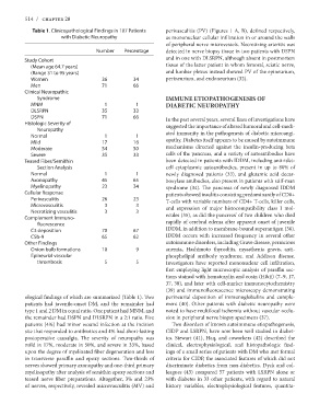
Table 1. Clinicopathological Findings in 107 Patients as mononuclear cellular infiltration in or around the walls
with Diabetic Neuropathy of peripheral nerve microvessels. Necrotizing arteritis was
detected in nerve biopsy tissue in two patients with DSPN
Number Percentage and in one with DLSRPN, although absent in postmortem
tissue of the latter patient in whom femoral, sciatic nerve,
Study Cohort 36 34 and lumbar plexus instead showed PV of the epineurium,
(Mean age 64.7 years) 71 66 perineurium, and endoneurium (32).
(Range 31 to 95 years)
Women 1 1 IMMUNE ETIOPATHOGENESIS OF
Men 35 33 DIABETIC NEUROPATHY
71 66
Clinical Neuropathic In the past several years, several lines of investigations have
Syndrome 1 1 suggested the importance of altered humoral and cell-medi-
17 16 ated immunity in the pathogenesis of diabetic microangi-
MNM 54 50 opathy. Diabetes itself appears to be caused by autoimmune
DLSRPN 35 33 mechanisms directed against the insulin-producing beta
DSPN cells of the pancreas, and a variety of autoantibodies have
Histologic Severity of 1 1 been detected in patients with IDDM, including anti-islet-
45 65 cell cytoplasmic autoantibodies, present in up to 80% of
Neuropathy 23 34 newly diagnosed patients (33), and glutamic acid decar-
Normal 26 23 boxylase antibodies, also present in patients with stiff-man
Mild syndrome (34). The pancreas of newly diagnosed IDDM
Moderate 3 3 patients showed insulitis consisting predominantly of CD8+
Severe 3 3 T-cells with variable numbers of CD4+ T-cells, killer cells,
Teased Fiber/Semithin and expression of major histocompatibility class I mol-
70 67 ecules (35), as did the pancreas’ of two children who died
Section Analysis 65 62 rapidly of cerebral edema after apparent onset of juvenile
Normal 10 9 IDDM, in addition to membrane-bound superantigen (36).
Axonopathy IDDM occurs with increased frequency in several other
Myelinopathy 5 5 autoimmune disorders, including Grave disease, pernicious
Cellular Response anemia, Hashimoto thyroditis, myasthenia gravis, anti-
Perivasculitis phospholipid antibody syndrome, and Addison disease.
Microvasculitis Investigators have reported mononuclear cell infiltration,
Necrotizing vasculitis first employing light microscopic analysis of paraffin sec-
Complement Immuno- tions stained with hematoxylin and eosin (H&E) (7–9, 17,
37, 38), and later with cell-marker immunocytochemistry
fluorescence (39) and immunofluorescence microscopy demonstrating
C3 deposition perineurial deposition of immunoglobulins and comple-
C5b-9 ment (40). Other patients with diabetic neuropathy were
Other Findings noted to have multifocal ischemia without vascular occlu-
Onion bulb formations sion in peripheral nerve biopsy specimens (37).
Epineurial vascular
Two disorders of known autoimmune etiopathogenesis,
thrombosis CIDP and LSRPN, have now been well studied in diabet-
ics. Stewart (41), Haq, and coworkers (42) described the
ological findings of which are summarized (Table 1). Two clinical, electrophysiological, and histopathologic find-
patients had juvenile-onset DM, and the remainder had ings of a small series of patients with DM who met formal
type 1 and 2 DM in equal ratio. One patient had MNM, and criteria for CIDP, the associated features of which did not
the remainder had DSPN and DLSRPN in a 2:1 ratio. Five discriminate diabetics from non-diabetics. Dyck and col-
patients (4%) had minor wound infection at the incision leagues (43) compared 57 patients with LSRPN alone or
site that responded to antibiotics and 4% had short-lasting with diabetes in 33 other patients, with regard to natural
postoperative causalgia. The severity of neuropathy was history variables, electrophysiological features, quantita-
mild in 17%, moderate in 50%, and severe in 33%, based
upon the degree of myelinated fiber degeneration and loss
in transverse paraffin and epoxy sections. Two-thirds of
nerves showed primary axonopathy and one-third primary
myelinopathy after analysis of semithin epoxy sections and
teased nerve fiber preparations. Altogether, 3% and 23%
of nerves, respectively, revealed microvasculitis (MV) and