
Page 64 - The Vasculitides, Volume 1: General Considerations and Systemic Vasculitis
P. 64
40 David S. Younger
caliber, from named cerebral vessels to small arteries and veins, was noted at postmortem
examination.
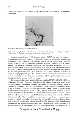
Reproduced from reference [4], with permission.
Figure 3. Radiographic features of cerebral vasculitis. Ectasia and beading in the M1 segment and lack
of flow in the A1 segment of the right anterior cerebral artery (arrow).
Salvarani and colleagues [187] diagnosed primary PCNSV in thirty-one patients by
histopathology and seventy patients by angiography, eighteen of whom had a granulomatous
inflammatory pattern, eight had a lymphocytic pattern, and five had an acute necrotizing
pattern. Headache was the commonest symptoms so noted overall in 63% of patients,
followed by abnormal cognition, hemiparesis and persistent neurological deficit. A
granulomatous pattern of inflammation was seen most often in those with altered cognition
and older age. There were no significant differences in survival when patients were stratified
by treatment (prednisone alone versus prednisone and cyclophosphamide) or method of
diagnosis (angiography or biopsy). Four manifestations at presentation were associated with
increased mortality rate including, focal neurological deficit, cognitive impairment, cerebral
infarction, and large cerebral vessel involvement.
The past decade has also revealed insights into childhood PACNS (cPACNS) [188], one
of the many childhood inflammatory brain diseases that affect small and large vessels as well
as, anti-N-methyl D-asparatate receptor encephalitis, anti-neuronal antibody mediated
diseases, and secondary CNS vasculitis in association with infection and rheumatic disease
[189] collectively known as childhood inflammatory brain disease. Angiographically-
negative small-vessel cPACNS (SVcPACNS) [190] lead to persistent headache, cognitive
decline, mood disorder, focal seizures and abnormal brain neuroimaging in children. Among
thirteen such children in whom detailed brain biopsy findings were available [191] the
inflammatory cell infiltrate located in intramural arterioles, capillaries, or venules in eleven
patients so studied, consisted predominantly of a mixture of lymphocytes and macrophages,
with occasional plasma cells, polymorphonuclear cells, and eosinophils. Granulomatous
inflammation and multinucleated giant cells were characteristically absent. Affected blood
vessels were found in leptomeninges, cortex, and subcortical white matter in seven of nine
Complimentary Contributor Copy