
Page 43 - The Vasculitides, Volume 1: General Considerations and Systemic Vasculitis
P. 43
Overview of Primary and Secondary Vasculitides 19
A
B
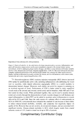
Reproduced from reference [4], with permission.
Figure 1. Giant cell arteritis. A. An early lesion of a large muscular artery, necrosis, inflammation, and
giant cell formation (single arrow) can be seen immediately adjacent to the internal elastic lamina
(arrowhead), which is undergoing degenerative changes, and there is some intimal proliferation (double
arrows) (stain, hematoxylin and eosin; original magnification, ×100). B. This more advanced lesion has
complete segmental destruction of the internal elastic lamina and virtually the entire media (arrows).
Marked intimal proliferation has nearly occluded the lumen, and few inflammatory cells remain (stain,
hematoxylin and eosin; original magnification, ×50).
18F- Fluorodeoxyglucose (FDG) positron emission tomography (PET) detects increased
FDG uptake by metabolically active cells, including inflammatory cells infiltrating the vessel
wall in vasculitis, while digital subtraction angiography (DSA) is a useful modality to
demonstrate luminal changes. Moreover, such studies can assist the surgeon in centering on
an involved segment of vessel. Performance of CDS is better suited to study superficial
vessels such as the internal and external carotid artery and its branches, while MR and CT are
best suited for deep vessels. When performed together, they can be used to monitor disease
extent and severity through the demonstration of early vascular changes in wall thickness and
mural inflammation, to which PET can be added to ascertain active inflammation in vessels
affected by GCA and TAK. Since the early reports of a salutary effect of corticosteroids on
GCA in 1950 [15], corticosteroids have remained the standard of care because of their ability
to reduce disease-related morbidity, mortality, and symptoms that negatively impact on
quality of life. However they are not curative, do not prevent relapses, and are associated with
significant toxicity. Disease-related morbidity in GCA which largely results from cranial
ischemic events or LVV, leads to visual loss in up to 20% of patients.
Complimentary Contributor Copy