
Page 366 - The Vasculitides, Volume 1: General Considerations and Systemic Vasculitis
P. 366
340 Sarah Conway and David S. Younger
more often in those with LVV aortic involvement than without (15% versus 54%).
Conversely, ischemic events were more common in those without than with evidence of LVV
aortic involvement by CT-imaging (54% vs. 22%). The authors postulated a spectrum of
GCA disease with cranial arteritis manifested by ocular symptoms, headache, and jaw
claudication at one end and predominant LVV at the other.
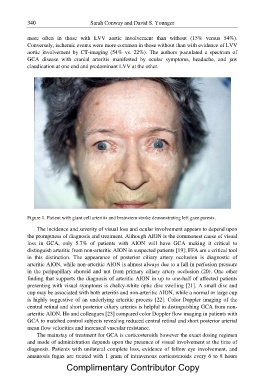
Figure 1. Patient with giant cell arteritis and brainstem stroke demonstrating left gaze paresis.
The incidence and severity of visual loss and ocular involvement appears to depend upon
the promptness of diagnosis and treatment. Although AION is the commonest cause of visual
loss in GCA, only 5.7% of patients with AION will have GCA making it critical to
distinguish arteritic from non-arteritic AION in suspected patients [19]; FFA are a critical tool
in this distinction. The appearance of posterior ciliary artery occlusion is diagnostic of
arteritic AION, while non-arteritic AION is almost always due to a fall in perfusion pressure
in the peripapillary choroid and not from primary ciliary artery occlusion (20). One other
finding that supports the diagnosis of arteritic AION in up to one-half of affected patients
presenting with visual symptoms is chalky-white optic disc swelling [21]. A small disc and
cup may be associated with both arteritis and non-arteritic AION, while a normal or large cup
is highly suggestive of an underlying arteritic process [22]. Color Doppler imaging of the
central retinal and short posterior ciliary arteries is helpful in distinguishing GCA from non-
arteritic AION. Ho and colleagues [23] compared color Doppler flow imaging in patients with
GCA to matched control subjects revealing reduced central retinal and short posterior arterial
mean flow velocities and increased vascular resistance.
The mainstay of treatment for GCA is corticosteroids however the exact dosing regimen
and mode of administration depends upon the presence of visual involvement at the time of
diagnosis. Patients with unilateral complete loss, evidence of fellow eye involvement, and
amaurosis fugax are treated with 1 gram of intravenous corticosteroids every 6 to 8 hours
Complimentary Contributor Copy