
Page 232 - The Vasculitides, Volume 1: General Considerations and Systemic Vasculitis
P. 232
Polyarteritis Nodosa 207
Cranial Nerve Manifestations
Albeit rare, involvement of the oculomotor, trochlear, abducens, facial, and
vestibulocochlear nerves are most often affected in PAN [45]. Effective treatment can lead to
partial or full recovery, which is less likely in the vestibulocochlear nerve [46]. There may be
vasculitis of the optic nerve, optic chiasm or occipital cortex. Cerebrospinal fluid is typically
normal.
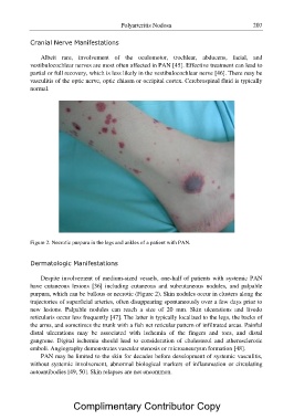
Figure 2. Necrotic purpura in the legs and ankles of a patient with PAN.
Dermatologic Manifestations
Despite involvement of medium-sized vessels, one-half of patients with systemic PAN
have cutaneous lesions [36] including cutaneous and subcutaneous nodules, and palpable
purpura, which can be bullous or necrotic (Figure 2). Skin nodules occur in clusters along the
trajectories of superficial arteries, often disappearing spontaneously over a few days prior to
new lesions. Palpable nodules can reach a size of 20 mm. Skin ulcerations and livedo
reticularis occur less frequently [47]. The latter is typically localized to the legs, the backs of
the arms, and sometimes the trunk with a fish net reticular pattern of infiltrated areas. Painful
distal ulcerations may be associated with ischemia of the fingers and toes, and distal
gangrene. Digital ischemia should lead to consideration of cholesterol and atherosclerotic
emboli. Angiography demonstrates vascular stenosis or microaneurysm formation [48].
PAN may be limited to the skin for decades before development of systemic vasculitis,
without systemic involvement, abnormal biological markers of inflammation or circulating
autoantibodies [49, 50]. Skin relapses are not uncommon.
Complimentary Contributor Copy