
Page 353 - Motor Disorders Third Edition
P. 353
THE HYPOTONIC INFANT / 335
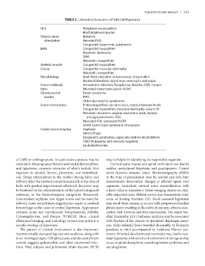
TABLE 2. Laboratory Evaluation of Infantile Hypotonia
NCS Peripheral neuropathies
Brachial plexus injuries
Tetanic nerve Botulism
stimulation Neonatal MG
Congenital myasthenic syndromes
EMG Congenital myopathies
Myotonic dystrophy
SMA
Metabolic myopathies
Skeletal muscle Congenital myopathies
biopsy Congenital muscular dystrophy
Metabolic myopathies
Microbiology Viral: Polio and other enteroviruses, Encephalitis
Bacterial: Botulism, diphtheria; meningitis and sepsis
Serum antibody Intrauterine infection: Toxoplasma, Rubella, CMV, Herpes
titers Neonatal myasthenia gravis: AChR
Chromosomal Down syndrome
studies PWS
Other dysmorphic syndromes
Serum chemistries Endocrinopathies: calcium levels, thyroid homone levels
Congenital myopathies, muscular dystrophy: serum CK
Metabolic disorders: organic and amino acids, lactate,
and pyruvate levels: ABG
Neonatal ADL: saturated VLCFA
Cranial neuroimaging Smith-Lemli-Opitz syndrome: cholesterol
Asphyxia
Hemorrhage
Dysgenetic syndromes, especially midline facial defects
CMD (Fukuyama and merosin-negative)
Leukodystrophies
of CMD or arthrogryposis. In utero toxin exposure may be may be helpful in identifying the responsible organism.
revealed in the pregnancy history and herald dysmorphism Cervical spine trauma and spinal cord injury can lead to
and hypotonia, common examples of which include fetal
exposure to alcohol, heroin, phenytoin, and trimethadi- sudden unexplained hypotonia and quadriparesis. Cranial
one. Drugs administered to the mother during labor and nerve function remains intact. Electromyography (EMG)
delivery affect the newborn most dramatically at the time of at the time of presentation may be normal and only later
birth with gradual improvement afterward. Recovery may demonstrates denervation changes at affected spinal root
be hastened by the administration of the opioid antagonist segments. Immediate cervical spine immobilization with
naloxone, or the benzodiazepine antagonist, flumazenil. a hard collar is mandatory before imaging studies in clini-
Concomitant asphyxia, low Apgar scores and the need for cally suspected cases. Skeletal survey may demonstrate other
delivery room resuscitation suggest prior sepsis or cerebral acute or healing fractures (23). Focal neonatal hypotonia
hemorrhage as the cause of central hypotonia. Appropriate may result from trauma as occurs with peripartum brachial
cultures, acute and convalescent Toxoplasmosis, Rubella, plexus injury resulting in flaccidity of one arm, often in asso-
Cytomegalovirus, and Herpes (TORCH) titers, cranial ciation with dystocia and fetal macrosomia. The upper bra-
ultrasound imaging, and toxicology screens may point to a chial plexopathy, Erb-Duchenne paralysis may be associated
specific etiology of hypotonia. with fracture of the clavicle or ipsilateral diaphragm paral-
ysis, while infantile lower brachial plexopathy or Klumpke
The pattern of clinical involvement is also important. paralysis, is often accompanied by ipsilateral Horner syn-
Asymmetrically decreased leg tone and weakness, along with drome. Inherited developmental anomalies may lead to neo-
fever, meningeal signs, CSF pleocytosis, and elevated protein natal hypotonia with selective involvement of the legs as may
content suggests poliomyelitis and other enteroviral infec- occur in spinal dysraphism, caudal regression syndrome, and
tions. Viral cultures and polymerase chain reaction (PCR) sacral agenesis.